What Does an Optometrist Do All Day? Glasses....Glasses....Glasses....or....

The Day in the Life of an Optometrist.png

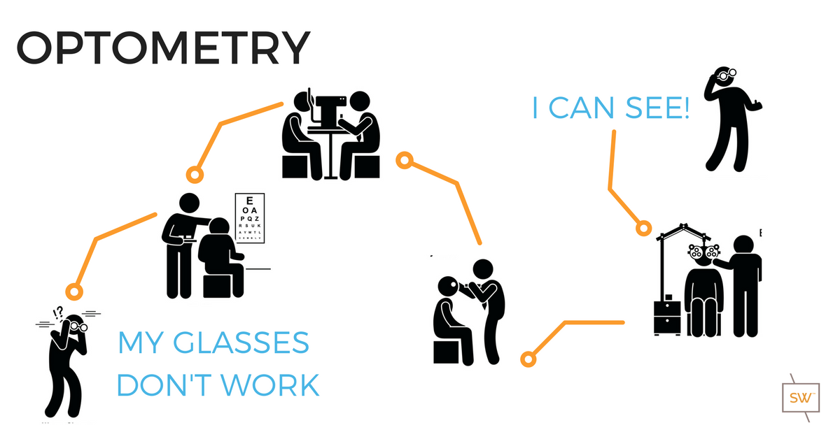
Have you ever wondered what an optometrists does all day?  

Most people know that if they are experiencing blurry vision, they can visit an optometrist who will provide them with a prescription for glasses or contacts.  But did you know that your optometrist does much more during your eye exam than merely prescribing glasses? And that not every cause of blurred vision can be fixed with glasses? That’s why every comprehensive eye exam also includes a thorough health check of your eyes.  Why do your eyes need a health check? And what exactly is your Optometrist looking for?

For us to be able to see, light must travel into and through the eye without difficulty, from front to back. When light enters your eyes, it must pass through several layers before reaching the back of the eye, where visual information is then sent to your brain via the optic nerve.

Your Optometrist needs to examine all of these anatomical layers to evaluate your eye's ability to receive and appreciate the light. To do this, they use an instrument called a "slit lamp", which utilizes a series of lenses and mirrors to magnify your eye, similar to a microscope.

The health of your eyes can change at any time and can be happening without you knowing it. The only way to be sure that your eyes are healthy is to have regular eye health checks. Most optometrists would recommend yearly eye health checks, just like your medical doctor and dentist do.

So what exactly is your optometrist looking for
 during these health checks?  

Let's start by reviewing the anatomy of your eyes!

  • The outermost layer of the eye is called the cornea. Many people confuse the cornea with the iris, which is the coloured part of the eye. The cornea is crystal clear and needs to stay that way to optimize the movement of light through its layers. The shape of the cornea is also partly responsible for determining the prescription of your glasses and contacts. For example, if your cornea is not perfectly spherical in shape, then you likely have some degree of astigmatism. Infections and scratches can cloud the cornea, causing a decrease in your vision. The cornea is protected by a tear film layer that is left behind after each blink you take. A compromised tear film can lead to dry eye symptoms such sandiness and grittiness on the front of your eye.

  • Surrounding the cornea on all sides, the “white of your eyes” is called the sclera. This area is a tough outer layer that acts like a shell to protect the eye from puncture. You might notice that the sclera appears red from time to time. This redness is due to the enlargement of blood vessels sitting on top of the sclera. These blood vessels are normally very thin and difficult to see but become enlarged as a reaction to irritation. This irritation can be caused by many things, including infection, inflammation and dryness.

  • Sitting behind the cornea is the iris or “coloured part of the eye”. The colour of the iris ranges from dark brown to pale blue depending on the amount of pigmentation present: a tremendous amount of pigment in brown eyes, a tiny bit of pigment in green eyes, and almost no pigment at all in blue eyes! In the middle of the iris, there appears to be a round black dot. However, this dot is actually a hole known as the pupil. You may have noticed that the size of the pupil changes throughout the day. This is normal. The pupil size will be large in dim lighting and small in bright lighting. It is the job of the iris to respond to the surrounding light levels with the appropriate pupil size for optimal vision. Inflammation of the iris, called iritis or uveitis, can affect the function of the iris, and lead to pain and light sensitivity.

  • Sitting behind the iris is the crystalline lens. "Crystalline" because it is crystal clear the day you are born. Just like the cornea, the lens needs to remain as transparent as possible to allow us to see the world around us. The lens is made up of collagen, is very flexible, and natural movements within the lens allow us to read. The lens becomes less flexible with time, and consequently, our reading ability gets weaker and weaker as we age. Decreased lens flexibility leads to the eventual need for reading glasses or progressive lenses. If the lens becomes cloudy or opacified in any way, whether by ageing, systemic conditions, or as a side effect of certain medications, it becomes what is known as a cataract. A cataract can significantly block the light travelling through the eyes and lead to a decrease in vision.

  • Sitting between the crystalline lens and the back surface of the eye is the vitreous humour. The vitreous is colourless and clear. It is a jelly-like tissue that is 99% water. The remaining 1% is made up of collagen, proteins, salts, and sugars. The vitreous provides protection to the eye as well as helps it to keep its spherical, or ball-shaped, appearance. With time, the collagen and proteins become "stringy", and the vitreous begins to liquefy. This creates symptoms of "floaters" which are more noticeable when looking at a white wall or a blue sky. Sometimes various health conditions necessitate the removal of the vitreous humour, and this is done through a process known as a "vitrectomy".

  • Once the light moves from the outside world, through the cornea, into the pupil and through the crystalline lens and vitreous humour, it finally hits the retina. The retina is where all the millions of receptors and nerves that convert the light into vision is located. Therefore it needs to be checked very carefully by your optometrist who will assess the macula, the optic nerve head, the blood vessels, and all other areas of the retina.

  • The macula is densely packed with photoreceptors and is responsible for your central, high-resolution vision. Damage to the macula can permanently affect your vision, and even cause blindness. Common causes of damage to the macula include smoking cigarettes and excessive sunlight exposure, which can lead to macular degeneration. The macula can also be damaged by uncontrolled diabetes. Permanent loss of vision due to damage to the macula can result in loss of one's driver's license and limited job opportunities.

  • The optic nerve is a collection site. Photoreceptors from the entire retina carry information to the optic nerve head, which is a direct line from the eye to the brain. Nerve damage from glaucoma is visible at the optic nerve head. Another important problem to rule out is papilledema, or swelling of the optic nerve head. This can cause headaches, blurred vision, and signal possible swelling within the brain itself.

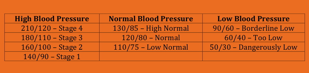
  • The blood vessels carry oxygenated blood and nutrients to the retina to maintain optimal function. Many systemic problems such as diabetes and high blood pressure can disrupt these blood vessels. It is important that your optometrist assess the blood vessels for leaks and proper blood flow.

Let's look at a few case examples, where your optometrist does a little more then you might think 

Patient with a corneal abrasion - visiting Dr. Holtz at Stonewire Optometry in Edmonton

Case Study 1:

Debbie Johnson goes to her optometrist, Dr. McKenzie, complaining of pain when opening and closing her left eye. Her left eye is also red and watery. Her right eye feels perfectly fine. Dr. McKenzie uses his slit lamp to examine her cornea. He notices a thin, whitish line on her cornea. It looks like a corneal abrasion, or scratch to the front of the eye. He then uses an orange dye called Sodium Fluorescien to help highlight the cornea and confirms that it is indeed a corneal abrasion

A corneal abrasion is an open wound to the eye and therefore can become infected if not treated. Dr. McKenzie starts Debbie on a course of antibiotics eye drops to fight any infection and to allow the cornea to heal itself. He may also prescribe lubrication eye drops to aid in comfort. It is important to discontinue any contact lens use while the cornea heals.


Gentlemen wth a cataract that requires care - visiting stonewire Optometry in Edmonton

Case Study 2:

Ben West goes to his optometrist, Dr. Lim, complaining of cloudy vision. He says it has been steadily getting worse over the last year and now he is noticing changes in his colour vision as well. Dr. Lim uses her slit lamp to evaluate his eyes. She notices a problem with the crystalline lens and recommends dilating Ben's eyes to get a better look at the lens as well as the retina to determine the cause of Ben's complaints. Once Ben's eyes are fully dilated from the diagnostic drops, Dr. Lim uses her slit lamp again. She notices that the crystalline lens has turned light brown in colour. No problems are found at the retina.

Ben's blurry vision and colour vision problem are due to cataracts and Ben will be sent to an Ophthalmologist for cataract surgery. Cataracts cause blurry vision that cannot be corrected by glasses and surgery becomes a necessity to continue daily tasks at the same level as before.


Patient with a macular hole - visiting Stonewire Optometry in Edmonton

Case Study 3:

John Jackson goes to his optometrist, Dr. McKenzie, complaining of warped central vision in his right eye. When he looks at something, like a tree, the trunk looks bent and distorted. Dr. McKenzie is very concerned about John's macula based on his complaint and recommends a dilated eye exam. 20 minutes after the dilation drops go in, Dr. McKenzie uses his slit lamp and a high plus lens to examine the macula of John's right eye. He sees a distortion of the macula and fluffy, yellow spots surrounding the macula.

John has signs and symptoms of macular degeneration. John needs to begin taking daily vitamins specially designed to aid the macula and fight off degeneration. If the degeneration continues to progress, John may need surgical intervention such as monthly injections. There are many risk factors that predispose someone to develop macular degeneration. These include excessive sun exposure, smoking, and a fair complexion.

Another common cause of John's symptoms is a macular hole. This can be caused by tugging of the vitreous body on the macula. This may or may not need surgical treatment, but if it does, John may need a vitrectomy, which is a removal of the vitreous body to alleviate the tugging on the macula.


Retinal Detachment.png

Case Study #4

Pamela Clark goes to her optometrist, Dr. Randhawa, complaining of a veil-like curtain over her left eye. She is also seeing flashes of light from the corner of her eye and increased number of floaters. Dr. Randhawa is very concerned about Pamela's retina based on her complaints and recommends a dilated eye exam. Once her eye is fully dilated, Dr. Randhawa uses her slit lamp to exam the retina of Pamela's left eye. Dr. Randhawa sees a small horseshoe-shaped tear in the outer retina. Pamela has a retinal tear, and emergent surgery is needed. Dr. Randhawa sends Pamela to the nearest hospital for surgery. Retinal tears and detachments can cause a permanent loss of vision and require immediate attention. It is important that anyone experiencing flashes or floaters be assessed right away by an optometrist.


Dry Watery Eye.png

Case Study 5:

Jane Fields goes to her optometrist, Dr. McKenzie, complaining of watery eyes throughout the day. Dr. McKenzie uses his slit lamp to exam the front surface of the eye and sees dry corneas. Jane's watery eyes are related to dry eye. This seemed counter-intuitive to Jane as her eyes are too wet, not too dry, but Dr. McKenzie explains to her that her cornea signals to the brain that it is dry and the brain responds by signalling to her glands to produce more tears. That is why a dry eye is a watery eye. Dry eye can be caused by many things including wind and a low humidity environment. It is helpful to wear sunglasses outside as well as to supplement with artificial tears.


Hopefully, this has given you an insight into the things your optometrist is looking for every time you come in for an eye exam.

Your eye health is essential to the function of your eye and needs to be examined on a regular basis. 


BOOK YOUR NEXT EYE EXAM IN EDMONTON
WITH A STONEWIRE OPTOMETRIST

Why Does My Eye Twitch?

A twitchy eye is generally considered a non-serious eye condition and is commonly experienced by patients. Medically, an eyelid twitch is referred to as‘Myokymia’.  The twitching sensation is the result of the sudden spasmodic contraction of the obicularious muscle, which is normally used for closing your eyelids. The twitching sensation most often effects the lower eyelid and occasionally can even place pressure on the eyeball itself causing the eye to twitch. Some patients are convinced that their eye is actually twitching, however, this is a vary rare phenomenon.  Symptoms from myokymia can last up to a month and can disappear as quickly as they began.

The most common causes of myokymia include:

  • Stress

  • Caffeine

  • Alcohol

  • Fatigue

  • Eye infection

  • Tourette Syndrome

  • Excessive exercise

  • Reduced potassium levels 

Although most eyelid twitches are not medically significant, it is always best to visit your optometrist or eye doctor to rule out any other more serious eye related diseases. Treatment for myokymia usually involves rest and relaxation, drinking plenty of water, and eating foods high in potassium. 

Fun Fact: Did you know that seeing your eye doctor for a condition like myokymia is now covered under Alberta Health Care.

If you're concerned about myokymia and your overall eye health, than book a routine eye health examination with one of our optometrists.

Book Your Next Eye Exam with Stonewire Optometry
Kingsway Mall | Edmonton | Alberta

Ophthalmology Referrals

If it’s determined during a routine or emergency eye examination that you require the care of an ophthalmologist, our eye doctors will help take care of the rest. The eye doctors at Kingsway Optometry work closely with many Edmonton based ophthalmologists to ensure that you have access to the appropriate health care provider.

Ophthalmology specialties available in Edmonton include:

  • Retina Specialists
  • Cornea Specialists
  • Glaucoma Specialists
  • Genetic Specialists
  • Oculoplastic Specialists
  • Pediatric Specialists
  • Strabismus Specialists
  • General Specialists

Ophthalmology Referral Process

  1. All referrals start off with a comprehensive eye health examination by one of our optometrists.
  2. Your optometrist will write a referral letter and call the ophthalmologists office to schedule your appointment.
  3. Depending on your eye condition and the urgency, patients will be seen right away, or up to 6 month. Most patients though are usually seen within 2-6 weeks.
  4. Our office will call you with your appointment information.
  5. Our office will also mail you a letter outlining your appointment information.  
  6. The ophthalmologists office will usually send a follow-up letter outlining the diagnosis, chosen treatment plan, and any follow-up care that may be required.


Have Your Eyes Examined Regularly

Of course! It seems obvious on the surface that one of the best things you can do to prevent vision loss from eye disease is to have your eyes checked regularly. Many eye diseases such as glaucoma, macular degeneration and diabetic retinopathy can be easily controlled if detected early enough, thereby preventing or limiting any loss of vision. It just makes sense that regular eye examinations would be the number 1 thing you could do to prevent vision loss. And yet still, it’s astounding just how few people regularly have their eyes checked.

In a study by the College of Optometrists in Vision Development, it was found that up to 25 percent of school-age children have some form of vision problem that may affect learning, but only a small percentage of children ever receive a comprehensive eye exam. With the new Eye See Eye Learn program by the Alberta Association of Optometrist, our clinic is working to change this by improving patient education and public awareness.

Even people with known eye problems aren’t getting eye exams. A study conducted at Duke University showed that only 70 to 90 percent of patients with glaucoma had a yearly check-up. Glaucoma is an eye disease that can slowly and silently cause blindness over a period of time. These patients often need to be followed at least two to three times a year. And yet a full 10 to 30 percent of them aren’t even going to the eye doctor yearly.

Similar numbers were found in patients with macular degeneration and diabetes. Only 65 to 80 percent of patients with macular degeneration have yearly eye exams and only 50 to 60 percent of patients with diabetes have them. Many of these people will very likely become legally blind from their untreated conditions. With the new medically necessary vision testing under Alberta Health Care your optometrist is able to follow these conditions (glaucoma, diabetes, and macular degeneration) at no charge to yourself.
The evidence speaks for itself – the number one recommendation is to make sure you have your eyes examined regularly!


Who Should I Go to For My Eye Exam?

There is often much confusion regarding whom to see for an eye exam – after all there are three different “O’s” that care for eyes in various ways. Who should you choose? To answer this question, let’s first define who the three “O’s” are:

  • Optometrists (O.D.’s) – A doctor of optometry receives his degree after four years of post graduate training in a college of optometry. Optometrists are licensed to perform comprehensive eye exams, prescribe glasses and contact lenses and to treat many types of eye disease including conjunctivitis (pink eye or red eye) and glaucoma. Optometrists typically do not perform surgery. Instead they receive additional training on understanding and treating the functional aspects of vision – how to help people see the best they can in their day to day lives.

  • Ophthalmologists – Ophthalmologists are medical doctors (M.D.’s) or osteopaths (D.O.’s) who take a three to five year residency in eye care after they receive their medical degree. In addition to providing comprehensive eye examinations, ophthalmologists are able to prescribe eye medications and perform surgery. Some ophthalmologists go on for additional training and become specialists in certain areas such as retinal, corneal, cataract and glaucoma treatment.

  • Opticians – Opticians are professionals specially trained to fit, fabricate and dispense eyeglasses. Many states also allow opticians to dispense contact lenses. Opticians do not perform comprehensive eye health examinations.

Which one should you choose for your routine comprehensive eye exams – an ophthalmologist or an optometrist? Obviously I may be biased, but I know today’s optometrists are well trained and skilled at detecting eye disease. If specialized medical or surgical treatment is ever needed, we will quickly arrange for you to see the right ophthalmologist who specializes in your particular eye care problem.


Meibominitis & Dry Eye

Meibomianitis is an inflammation of the oil glands or meibomian glands along your upper and lower eyelid margins. These oil glands produce the lipid or fat layer of your tear film, which coats the surface of your eye. The purpose of this lipid layer is to help prevent the water layer below from evaporating. When these meibomian glands or oil glands become inflamed, they no longer produce nice smooth clean oil. The oil can come out bubbly or thick, thus improperly coating the front of the eye.

Meibomianitis typically does not cause serious visual problems, but it can cause numerous comfort and cosmetic issues. Meibomianitis can cause your eyes to dry out, burn, itch, and water. All of which can lead to reduced vision, blurred vision, vision that fluctuates or vision that clears up with strong blinks. Meibomianitis can also cause your eyelids to swell or thicken and turn red, which can eventually lead to madarosis (eyelashes falling out). It can also lead to external hordeolums more commonly known as styes, which are simply clogged oil glands that have become inflamed.

TREATMENT OR PREVENTION:

The treatment for meibomianitis is fairly straight forward, and we encourage all of our patients to perform this treatment on a daily basis, regardless of whether or not they have meibomianitis.

Steps:

  • Prior to getting in the shower in the morning, take a washcloth and heat it up under the tap with warmwater. Close your eyes, and lightly hold the warm washcloth over your eyes. Hold the cloth there until it cools off. Repeat this process for approximately 2 minutes.
  • Than, lightly rub your eyelids with the wash cloth to force out all the old oil
  • If you eyelids are really swollen than in the shower, take your washcloth and apply a small amount of baby shampoo to it (baby shampoo is used so that you eyes won’t burn, if they actually come in contact with the soap). Foam it up. Than lightly scrub you eyelid margins to remove any oil build up. Ensure that you thoroughly rinse away any soap residue.

Occasionally meibomianitis can get very bad, and medical therapy may be required. If this is the case, your optometrist or ophthalmologist may provide you with a topical antibiotic ointment. In some situations, oral antibiotics may also be used. 

To learn more about meibomianitis or to find out if meibomianitis is the cause of your vision problems, please contact one of our eye doctors and book and eye exam. 


High Blood Pressure and Your Eyes!

Having high blood pressure or hypertension (HTN) can have a serious impact on a persons overall well being along with the overall health of their eyes. It is estimated that approximately 22% of Canadians have hight blood pressure, but that only about half actually receive treatment for it. High blood pressure is considered to be a silent killer, as it often has no symptoms at all.

Elevated blood pressure is defined as having a systolic pressure of greater than 140mmHg or a diastolic pressure of more than 90mmHg. In the vast majority of people, there is no specific cause for their elevated blood pressure, but studies show that high blood pressure is more common in African Americans than in Caucasians and more common in the elderly. High blood pressure is also more likely to develop in people suffering from obesity or diabetes.


If blood pressure is aloud to remain elevated for any length of time, it can start to impact your health

The heart may become larger or weaker, which may lead to heart failure. Heart failure is when the heart cannot pump enough blood throughout the body

Aneurysms can form in blood vessels. Aneurysms are abnormal bulges or ballooning of an artery wall. Common locations are the brain, legs, stomach and spleen

Blood vessels in the kidneys can start to narrow, leading to kidney failure

Blood vessels can start to narrow throughout the body, which can lead to heart attacks, stroke, or kidney failure.

In men, high blood pressure can also cause erectile dysfunction

Blood vessels in the eyes can burst or bleed, which can lead to vision changes or blindness.

GENERAL SYMPTOMS:

  • No symptoms
  • Headaches or Morning Headaches
  • Tinnitus (ringing in the ears)
  • Dizziness
  • Nose Bleeds
  • Chest Pain
  • Flushed or Pale Skin
  • Nausea/Vomiting

EYE SYMPTOMS:

  • Papilloedema (swelling of the optic nerve- can cause pain with eye movements or loss of colour vision)
  • Blurred Vision
  • Double Vision

TREATMENTS:

  • Lifestyle 
  • Lose Weigh
  • Quit Smoking
  • Eat a healthy diet
  • Reduce Salt intake
  • Reduce alcohol consumption
  • Medications

**There are numerous types of high blood pressure medications on the market today. Only your family physician and yourself can determine the best type of medication for you.

For more information on High Blood Pressure or Hypertension, please visit the Heart and Stroke Foundation. If you are concerned that High Blood Pressure may be affecting your vision, please see your optometrist. Your optometrist has the knowledge and skill to work along with your family physician when managing your blood pressure.