
OPTOMAP - ULTRA-WIDEFIELD RETINAL IMAGING
Optomap ultra-widefield digital imaging is an exciting imaging technology that allows both you and your optometrists to see a 200-degree picture or image of the inside of your eye or about 80% of the retina.
This ultra-widefield digital image provides a ‘map’ that helps to guide your optometrist during the eye exam towards areas of concern. Your eye doctor can also share these images with you, becoming an invaluable tool in patient education. You can read more about this in our blog post: You see what we see; the benefits of Optomap
"We introduced Optomap digital retinal imaging technology into Stonewire Optometry because we found it helped provide our patients with peace of mind, knowing that their eyes were healthy. While clinically it serves as a great education tool and an improved way to monitor areas of concern" Dr. Ross McKenzie, OD.

California Autofluorescence Optomap

Stonewire uses the California af Optomap model which images the eye faster than previous generations, allowing younger and older patients to be imaged in just seconds.
The California af was the first Optomap to incorporate both composite colour and autofluorescence digital imaging. You can see an example below of the difference in the digital images. The autofluorescence digital image provides us with another tool in trying to determine the cause of underlying vision complaints. It can help our eye doctors uncover problems that would previously go undiagnosed or explained.
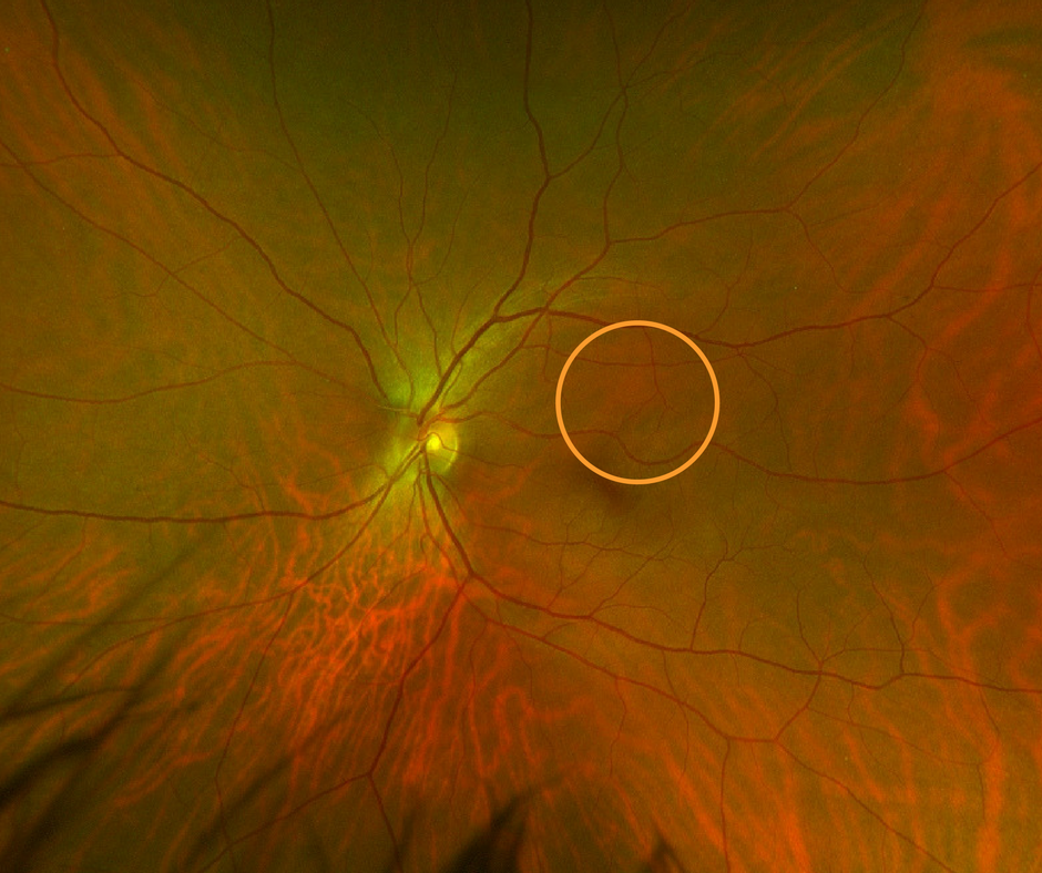
Composite Colour Imaging
Composite colour digital imaging uses multiple lasers to image the back of the eye. These lasers penetrate the layers of the retina to different depths, allowing your optometrist to visualize and located the layers of the retina more easily. This aids in the detection, diagnosing and monitoring of various eye conditions. The Green (532nm) laser visualizes the sensory retina to the RPE layer. The Red (635nm) laser looks deeper between the RPE to choroid. When combined, they produce the digital image you see above, which can be used to help your optometrist explain your eye health.

Autofluorescence Imaging
The auto-fluorescence (AF) mode on the Optos California model helps our optometrists monitor retinal conditions by showing abnormalities or metabolic changes within the retina that may not be visible during standard digital retinal photography or in a dilated retinal exam. This test is valuable in patients with conditions such as diabetic retinopathy, age-related macular degeneration or hydroxychloroquine toxicity because it can help determine if the affected area is in an active or stable phase. This non-invasive test can regularly be performed on patients of all ages.
Optomap digital images are exciting and educational for patients to look at
and offer our optometrists further insight into your eye health.
The digital images don’t replace a dilated retinal exam, they just make it better.

Although optomap California provides our optometrists with an ultra-widefield view of your retina, it does not replace the need for a regular dilated retinal exam. Patients with conditions such as diabetes, hypertension, glaucoma, and age-related macular degeneration still require this procedure annually, as it's still considered the standard of care. A dilated exam provides more detail to your optometrist by understanding elevations and depressions, clarity of the eye, and a more dynamic view of the retina.
Optomap ultra-widefield images are simply a tool our optometrists use to help uncover and monitor eye diseases during a routine eye exam. The test is especially useful in asymptomatic patients or who may not receive regular dilated retinal exams. They also provide a digital record which we can use to monitor eye conditions year to year.
These Ultra-widefield images also provide an excellent way to educate our patients on their eye conditions or ensure their eyes are healthy.
YOU SEE WHAT WE SEE
If something is detected during an optomap exam, your optometrist will need further testing. Testing may include visual field analysis, OCT retinal imaging, ultra-widefield autofluorescence photography, or digital retinal photography of the central retinal pole.
Stonewire Optometry is the only eye care clinic in Kingsway Mall offering this unique imaging service. If you’re looking for an in-depth eye exam with the latest equipment, please book your eye exam online today.
Cost: We include OPTOMAP in all comprehensive regular pay Adult Eye Exams*
*Images may not be captured on all patients because of pupil size or mobility issues.
*If additional images are required to monitor your condition, they may be covered under Alberta Health Care. If you do not have Alberta Health Care, the cost is $60.00 when not part of a routine eye exam.

Is Optomap Imaging safe for children?
Yes. Optomap images are safe to take on children. Eye disease can affect children just like it affects adults. Quality routine eye care can provide peace of mind, and help parents better understand their child's eye health.
How new is Optomap technology?
Optomap has been around since 1999 and is currently being used in about 8700 optometry and ophthalmology offices around the world.
Is optomap as comprehensive as the old way I had my eye examined?
Optomap is additive to your eye exam. It gives both you and your eye doctor and view that was previous unavailable. It adds to you doctor's ability to diagnose the health of your eye, and it better allows you to understand what is happening. Imaging also allows us to better monitor your eye health for changes, good or bad.

